Testimonials:
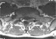
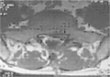
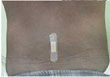
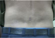
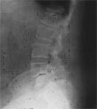
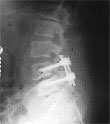
An elderly man presented with severe radiating pain in his right leg. The MRI showed a large disc prolapsed at L4/5 level. The patient was unable to walk straight before operation. Within 6 hours after endoscopic surgery, patient could walk straight without any support. Before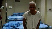 After
After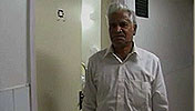 |
Endoscopic Surgery for Disc Prolaps
Slipped disc or Sciatica is a common problem, which affects approximately 1% of the populationat any given time. It usually affects people between the ages of 30 to 50 years. It is more common in males and frequently occurs in people whose occupation involves frequent bending and lifting.
The slipped disc typically causes low back pain, which travels to buttock and further down to back of thigh and calf muscles. This leg pain is also called as Sciatica because it radiates along distribution of sciatic nerve. This pain increases on sneezing or straining, walking etc. Pain may be accompanied by numbness or weakness in leg muscles and in severe situations bladder and bowel disturbances.
In recent years, MRI scanning is becoming available more easily and is the preferred diagnostic test for spinal problems. MRI scan can clearly show the site and amount of disc prolapse and the nerve compression.
Majority of patients respond to the non-surgical treatment. Approximately 80% will not require any operation. Non-surgical treatment is given in the form of bed rest, medicines to reduce pain and swelling. Physiotherapy is gradually started.
Surgery is required in 10-20% of patients whose pain persists after conservative treatment or who have paralysis or bladder and bowel dysfunction from beginning. Traditionally, surgery for the slipped disc is performed by about 4-5cms. incision in the back and then using a microscope, small portion of bone is removed and then disc is removed.
But now-a-days operation for slipped disc can be performed through a tiny (about 1.5 cms) incision in the back with the help of an endoscope. Patients need not be hospitalized and they can go back home in the same evening. Operation can be performed without general anesthesia. A band-aid is applied on the incision after surgery. Patients are very comfortable after operation and they can start walking or going to toilet same day. Post-operative period is less painful. Patients can go back to their work in a few days.
Endoscopic Surgery for Pituitary Tumours
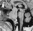
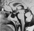
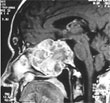
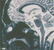
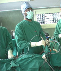 Pituitary tumours are mostly non-malignant brain tumours and account for approximately 10% of all brain tumours. Their incidence in general population is about 1 out of 10.000 people. These tumours arise from the master hormone gland of the body i.e. pituitary gland. Pituitary tumours are mostly non-malignant brain tumours and account for approximately 10% of all brain tumours. Their incidence in general population is about 1 out of 10.000 people. These tumours arise from the master hormone gland of the body i.e. pituitary gland.
Patients commonly present with the vision problem, hormonal disturbances or headache. Hormonal problems can be in the form of irregular or absence of menses, discharge from breasts, excessive growth of facial features or body, growth of hair over body, decreased sexual drive and impotence in men etc.
With conventional techniques, pituitary tumours are removed either by performing a craniotomy or transsphenoidally in a more traumatic way.
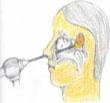
But now these tumours can now be removed through nose with help of a 4-mm. Endoscope in a least traumatic way. No incision is given on the face or head. There is no dissection in nose therefore post-operative nasal packing is not required. Patients are very comfortable after the operation and can usually go home next day after operation.
Endoscopic Surgery for Hydrocephalus
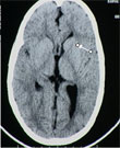 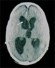 Hydrocephalus is one of the most commonly treated diseases by neurosurgeons. It means accumulation of excessive fluid (Cerebro-spinal fluid) in the brain. It commonly occurs due to the obstruction in the flow of fluid in the brain, sometimes it can occur due to poor absorption of fluid and rarely due to the overproduction of fluid. In newborn babies it presents with progressive enlargement of head size while at other ages it can present with headache, vision problems, or deteriorating brain functions like diminishing memory, altered consciousness, walking problems, poor control on sphincters etc. Hydrocephalus is one of the most commonly treated diseases by neurosurgeons. It means accumulation of excessive fluid (Cerebro-spinal fluid) in the brain. It commonly occurs due to the obstruction in the flow of fluid in the brain, sometimes it can occur due to poor absorption of fluid and rarely due to the overproduction of fluid. In newborn babies it presents with progressive enlargement of head size while at other ages it can present with headache, vision problems, or deteriorating brain functions like diminishing memory, altered consciousness, walking problems, poor control on sphincters etc.
For decades the standard treatment has been the placement of a shunt tube from brain to the abdomen to bypass the fluid. But the problems with shunt are that it gets blocked frequently or it can get infected, in either case frequent re-operations are required. Now with the help of an endoscope, an internal bypass of fluid can be established. This procedure, third ventriculostomy, is performed through a small hole in the skull bone and patient can go home same day or next day after surgery. This procedure can completely cure the problem.
Endoscopic Surgery for Brain Tumours
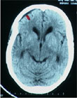 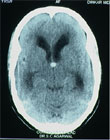 Many brain tumours and tumour like conditions like colloid cyst, intra ventricular cysticercus, arachnoid cyst; intraventricular tumours can be treated endoscopically through small hole in the skull. Colloid cysts are, one of such examples, found in third ventricles and they block the CSF pathway to produce hydrocephalus. These cysts can be removed endoscopically through a small hole in the skull and cure the patient. Many brain tumours and tumour like conditions like colloid cyst, intra ventricular cysticercus, arachnoid cyst; intraventricular tumours can be treated endoscopically through small hole in the skull. Colloid cysts are, one of such examples, found in third ventricles and they block the CSF pathway to produce hydrocephalus. These cysts can be removed endoscopically through a small hole in the skull and cure the patient.
|
Endoscopic Surgery for CSF Rhinorrhea
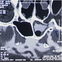 CSF Rhinorrhea is the leakage of brain fluid through the nose. It can occur after head injury, after brain or nasal surgery or sometimes spontaneously. Majority of patients require operation to stop leakage. Earlier, a major operation was performed by craniotomy in the skull. Patients had to stay in the hospital for a longer time and they used to loose their sense of smell. But now with the help of a 4-mm. endoscope, these leaks can be stopped permanently through the nose. Patients don't loose their sense of smell and can go back home early. CSF Rhinorrhea is the leakage of brain fluid through the nose. It can occur after head injury, after brain or nasal surgery or sometimes spontaneously. Majority of patients require operation to stop leakage. Earlier, a major operation was performed by craniotomy in the skull. Patients had to stay in the hospital for a longer time and they used to loose their sense of smell. But now with the help of a 4-mm. endoscope, these leaks can be stopped permanently through the nose. Patients don't loose their sense of smell and can go back home early.
|
Stereotactic Surgery for Brain Hemorrhage
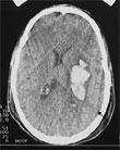 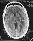 Brain hemorrhage, which is a very serious condition, often presents with paralysis or unconsciousness. In many cases it can be treated by aspiration of hemorrhage through a small hole in the skull under local anaesthesia. Patients usually show a remarkable recovery after operation Brain hemorrhage, which is a very serious condition, often presents with paralysis or unconsciousness. In many cases it can be treated by aspiration of hemorrhage through a small hole in the skull under local anaesthesia. Patients usually show a remarkable recovery after operation
|
|
Stereotactic Surgery for Brain Tumor
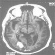 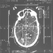 Many deep seated or inaccessible brain tumours may be biopsied through a small hole under local anesthesia. Many deep seated or inaccessible brain tumours may be biopsied through a small hole under local anesthesia.
|
|
|
Microsurgery for Brain Tumors
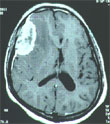
 Brain tumour surgery-- the very words strike fear in the heart of anyone threatened by one. It once was considered one of the most frightful events that could occur. Today, however, with improving technology, patients and families can look to the future with considerably more hope. Historically, patients undergoing brain surgery would inevitably be faced with paralysis, coma, long hospitalization, staying away from work for long time. The general misconception of depicting the neurosurgical patient with his head shaved, wrapped in an extensive dressing, often in a coma for days only to make a miraculous, but unfortunate and incomplete recovery. But with
 |
Microsurgery for Spinal Tumours
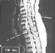 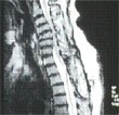
Spinal tumours are removed under microscope with no trauma to surrounding spinal cord/ nerve roots.
|
For Appointment Call :
011-25854855
011-32903273
or Email us at:
mail@drsatnamchabra.com
|
Photo Gallery : |
|